Previously, we have given the procedure to make a simple stain, one that is used to identify colony and morphology of a given bacteria .
Today, we were able to discover the colony and morphology of our unknown because our pure culture had individual colonies in it! Here our some pictures of the unknown:
The colony was determined as predicted! The colonies in this picture are obviously circular. They had a cream tint to them and covered the entire surface. They were opaque and raised off the agar medium.
Even with the colony and morphology identification, it is still impossible to know the exact identity of our bacteria without doing deeper investigation! Our next step was to do a Gram Stain to identify the composition of our bacteria's cell wall.
To prepare a Gram Stain:
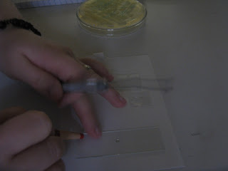
First, to create our stain, we followed the procedure used to make a simple stain: smearing, drying, and heat-fixing the bacteria onto a slide. The full procedure for this can be found in our previous blog, "Staining Bacteria: Preparing a Smear and Preparing an Unknown Sample."
The Gram Stain, however, is more complex. It involves the use of three stains: Crystal Violet, Gram's Iodine, and Safranin.
The initial stain used was Crystal Violet; we covered the fixed smear with this stain for 20 seconds while the slide was suspended over the sink on a slide drying rack.
After this, we rinsed the slide with water and covered the smear with dye again. However, this time we used Gram's Iodine, keeping the smear covered for 1 minute. After rinsing a second time, we used 95% enthanol (labeled EtOH above) to decolorize the sample. We held the slide at a 45 degree angle until the color stopped running off the smear. It is important to rinse the slide with water immediately after the decolorizing process is complete so color doesn't continue to fade.
After this, the last dye was used: Safranin. This dye was left on the slide for 1 minute before rinsing for the last time and blotting the slide dry with Bibulous paper.
The results!
The Environmental Sample:
This sample is Gram-negative. We know this because it took up the Safranin dye. Because of the small peptidoglycan layer in a Gram-negative bacteria, the purple dyes were unable to stay fixed in the cell wall when the ethanol (decolorizing agent) was applied. We predict that this bacteria produces endotoxins because the cell walls of Gram-negative bacteria have LPS in them. This type of bacteria is harmful when it gets inside the body because it activates the immune system of the host. When this cell is lysed, the LPS causes coagulation and much more. REMEMBER: this bacteria was found on the bathroom door! If someone were to touch this door without washing their hands and picked up enough of this bacteria, they could sick!
The Unknown "F":
This sample is Gram-positive. This stain took up the Crystal Violet and Gram's Iodine stains because of its thick peptidoglycan layer. The cell wall of this bacteria also has teichoic and lipoteichoic acids in it because these acids are found only in Gram-positive bacteria. These acids stabilize the cell.
As seen in the two samples above, both of our bacteria are small, cocci bacteria. This makes sense because as seen in the previous blog, both samples were also non-motile. Cocci bacteria are generally non-motile.
We're getting closer and closer to identifying our bacteria! What will they be? Harmful? Contagious? Stay posted to find out!


No comments:
Post a Comment